Pick an area on the figure and you will see what lies underneath it: which muscle it is, what it does, where it usually refers pain and what hands actually do with it during a session. This is a map for a conversation, not a diagnosis.
What this atlas is for
So that we are talking about the same place
The most common description of pain at a first session is a palm circling over the back and the words “somewhere around here”. That is a perfectly normal place to start. But the more precisely you name the spot and the circumstances, the less time goes into searching and the more into the work itself.
Pain almost never comes from the point you are jabbing with your finger. It comes from an area that has borders, a function and a characteristic way of letting you down: one muscle aches only under load, another the opposite — after an hour of sitting still, a third stays completely silent while the “pain” turns up a hand’s width to the side. The atlas shows these areas on anatomical 3D renders: a figure without skin, musculature visible, none of those textbook diagrams that are attached to nothing.
The practical benefit is simple. You come in and say not “my back”, but “it pulls along the spine between the shoulder blades, worse towards the evening, gone after sleep”. That is already a working description: it is clear where to begin palpating, which position to put you in on the table, how much pressure to hold and which movements to test before and after. At a first session that saves about fifteen minutes — roughly a quarter of a short session.
The second benefit is that you stop being afraid of your own body. When “that strange thing in my lower back” gets a name, borders and an explanation, the anxiety drops noticeably, and that affects muscle tone directly. And you start noticing connections: that three days behind the wheel speaks up in one place, and the gym in a completely different one.
Where the muscle begins and ends — that is, how far hands can honestly reach, and where another area starts
What the muscle does in movement, and which posture keeps it working for hours with nothing to show for it
Where it usually refers pain — and why “it hurts here” does not mean “the cause is here”
How it is normally worked: direction, pressure, how much of a session it takes
When it is left alone entirely — a fresh injury, inflammation, a symptom that does not add up
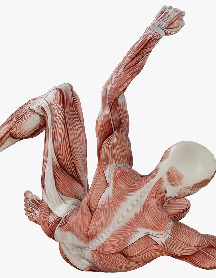
A twisted position separates the layers of the back: on top the trapezius (m. trapezius) and the latissimus dorsi (m. latissimus dorsi), beneath them along the spine the erector spinae (m. erector spinae), lower down the glutes. That is exactly how these areas are opened up on the table — with rotation, not with force.
Trapezius (m. trapezius)
Latissimus dorsi (m. latissimus dorsi)
Erector spinae (m. erector spinae)
Glutes (mm. glutei)
Muscle atlas: what exactly hurts, and why
Hover over a marker on the map and you will see where the muscle sits, what it does, how its overload shows up and what deep tissue massage does with it. This is an anatomy reference, not a diagnosis: pain is good at misleading you, and the spot that aches is often only an address rather than the source.
The base of the interactive atlas: a map of the back of the body marking the muscles that most often need deep work.
Trapezius (m. trapezius)
Deltoid (m. deltoideus)
Latissimus dorsi (m. latissimus dorsi)
Erector spinae (m. erector spinae)
Thoracolumbar fascia (fascia thoracolumbalis)
Glutes (mm. glutei)
Hamstrings (mm. ischiocrurales)
Gastrocnemius (m. gastrocnemius)
Upper trapezius
m. trapezius (pars descendens)Neck and shoulders
Where it sits
Runs from the occipital bone and the cervical vertebrae to the outer third of the collarbone and the acromion — the very “shelf” between the neck and the shoulder.
What it does
Lifts the shoulder girdle, helps tilt and turn the head, and carries the weight of the freely hanging arm.
How it hurts
Heaviness along the top edge of the shoulder, often spreading up the side of the neck — towards the temple and behind the ear.
What overloads it
A bag on one shoulder, armrests set too high, an unsupported mouse hand, a phone wedged between ear and shoulder, a draught of cold air from the air conditioning.
How we work on it
Slow cross-fibre pressure and a hold on the dense spot for 30-60 seconds, for as long as your breathing stays even. Your head rests in my hands the whole time — the muscle must not be working if it is going to let go.
Middle and lower trapezius, rhomboids
m. trapezius (pars transversa et ascendens), mm. rhomboideiBetween the shoulder blades
Where it sits
Between the spine and the inner border of the shoulder blade: from the spinous processes to the spine of the scapula and below it, under the inferior angle.
What it does
They draw the shoulder blades in towards the spine and pull them down, keeping the scapula held against the rib cage.
How it hurts
Burning between the shoulder blade and the spine by the end of the working day, and a nagging urge to crack your back — followed by a short spell of relief.
What overloads it
Hours with rounded shoulders, a laptop below eye level, a steering wheel with the arms stretched out to it, working with two monitors placed off to the sides.
How we work on it
An honest caveat belongs here: the muscles between the shoulder blades are more often overloaded while lengthened, and it is the opposite side — the pectorals — that is short. So we work both along the edge of the shoulder blade and on the front of the chest; otherwise the relief lasts an hour.
Suboccipital muscles
mm. suboccipitalesBase of the skull
Where it sits
Four pairs of short muscles between the occipital bone and the top two cervical vertebrae, under a thick layer of trapezius.
What it does
Fine-tuning the position of the head: micro-nods and micro-turns. They are closely tied to eye movement — the gaze leads, the neck answers.
How it hurts
A pressing headache that runs from the back of the head like a band over the crown to the eye, and a sense of a heavy head by evening.
What overloads it
A day spent looking slightly up at a screen, reading lying down, a pillow that is too high, holding your gaze on one point for long stretches.
How we work on it
Finger pressure held along the base of the skull: the head rests in my hands, the pressure moderate and static. No jerks and no fast rotations of the neck — precision counts here, not force.
Scalenes and sternocleidomastoid
mm. scaleni, m. sternocleidomastoideusSide and front of the neck
Where it sits
The scalenes run from the cervical vertebrae to the top two ribs; the SCM is a cord from the mastoid process to the breastbone and the collarbone.
What it does
They tilt and turn the head and lift the upper ribs on the inhale — which is why they tire first in people who breathe into the chest.
How it hurts
The SCM refers pain to the forehead and behind the eye; the scalenes give heaviness and tingling down the outer side of the arm.
What overloads it
Shallow chest breathing, a lingering cough, a high pillow, the habit of carrying a child or a bag always on the same side.
How we work on it
Layer-by-layer work with a soft pincer grip on the SCM and precise pressure on the scalenes. Blood vessels and a nerve bundle run right alongside, so the pace is slow and feedback is essential: numbness or a shooting sensation into the arm is a signal to stop and see a doctor. On the front of the neck, where the carotid arteries run, no pressure is applied at all.
Deltoid and rotator cuff
m. deltoideus; rotator cuff: m. supraspinatus, m. infraspinatus, m. teres minor, m. subscapularisShoulder joint
Where it sits
The deltoid caps the joint from the outside; the cuff lies deeper — above the spine of the scapula, below it and on the inner surface of the shoulder blade.
What it does
The deltoid raises and abducts the arm; the cuff keeps the head of the humerus centred in the joint throughout the whole movement.
How it hurts
A dull ache down the outside of the upper arm, trouble lifting the arm out to the side or reaching behind your back; at night it gets in the way of lying on that side.
What overloads it
Pressing and pull-ups with nothing for the back of the cuff, long stretches of work with the arms overhead, swimming, painting a ceiling.
How we work on it
Work on supraspinatus and infraspinatus through the shoulder blade, and a careful approach to the edge of the cuff with the arm abducted. Massage takes care of the muscular part of the problem, but it does not replace an examination: if a tendon tear is suspected, you need a doctor and imaging.
Latissimus dorsi
m. latissimus dorsiSide of the back
Where it sits
It starts broadly — from the thoracolumbar fascia, the lower thoracic vertebrae, the crest of the hip bone and the lower ribs — and converges into a narrow tendon at the humerus.
What it does
It draws the arm in and down, pulls the body up in a pull-up and links the pelvis to the shoulder: the only muscle thrown across the entire back.
How it hurts
Restriction when raising the arm overhead (“I cannot reach the top shelf”), a pulling ache along the side and under the lower angle of the shoulder blade.
What overloads it
Rowing, pull-ups, climbing, long spells working with the arms out in front, sleeping on your side with the arm tucked under.
How we work on it
Taking hold of the muscle near the armpit and pressing slowly along the fibres down towards the lower back, often combined with passively raising the arm. It is exactly the combination of pressure and movement that makes the difference here.
Erector spinae
m. erector spinaeAlong the spine
Where it sits
Three columns — iliocostalis, longissimus and spinalis — run from the sacrum up to the back of the head as two ridges either side of the spinous processes.
What it does
They hold the trunk upright and brake the forward bend. Sitting without leaning on a backrest keeps them working almost without a pause.
How it hurts
A diffuse fatigue along the lower and mid back, the feeling that your back “gets tired of standing”, stiffness in the morning.
What overloads it
A day spent sitting, a long drive, work done bent over, standing still in one spot for a long time.
How we work on it
Long forearm passes along the ridges and cross-fibre separation of the layers on the dense spots. Pressure goes into the muscle only, set back from the midline: the spinous processes are never pressed on.
Quadratus lumborum
m. quadratus lumborumDeep lower back
Where it sits
It lies deep, between the 12th rib and the crest of the hip bone, underneath the erector spinae. It is reached from the side, with you lying on your side.
What it does
It bends the trunk sideways, draws the pelvis up towards the rib, stabilizes the lower back with every step and anchors the 12th rib on the inhale.
How it hurts
One-sided deep pain in the lower back: worse when getting up out of a chair and when coughing, referring into the buttock and towards the crest of the pelvis.
What overloads it
Sitting lopsided (a wallet in the back pocket), a difference in leg length, lifting something heavy with a twist, driving with one foot permanently on the pedal.
How we work on it
Approached from the side, past the outer edge of the erectors, layer by layer and on the exhale. This is the usual reason rubbing the middle of the back leads nowhere: the lower back is what hurts, but the overloaded muscle sits beneath it.
Thoracolumbar fascia
fascia thoracolumbalisLumbosacral diamond
Where it sits
A dense diamond of connective tissue over the sacrum and the lower back, into which the latissimus, the glutes and the abdominal muscles weave.
What it does
It passes force from one half of the body to the other — from the right glute to the left latissimus — and works as a natural belt under load.
How it hurts
Not a point but a quality: a “stiff” lower back, the feeling of a tightened corset, stiffness after a long spell without moving.
What overloads it
Static positions, glutes that do little of the work, monotonous loading, guarded movement habits left over from an old injury.
How we work on it
Slow pressure with a long vector and a hold: fascia responds to time and warmth, not to force. A fast, hard pass only earns you a protective tightening in return.
Gluteal muscles
m. gluteus maximus, m. gluteus medius, m. gluteus minimusPelvis
Where it sits
Gluteus maximus gives the buttock its volume; medius and minimus sit higher and further out, closer to the crest of the hip bone, one beneath the other.
What it does
Maximus extends the hip — walking, climbing stairs, standing up. Medius keeps the pelvis level while you stand on one leg.
How it hurts
Gluteus medius refers pain along the crest and into the outer part of the buttock and is easily mistaken for “the lower back”; when you walk, the pelvis drops to one side. Minimus lies beneath it and refers differently — down the outside and the back of the thigh, which is why it is regularly taken for “a pinched nerve”.
What overloads it
Desk work, running on a cambered roadside always in the same direction, standing for long stretches with the weight on one leg, carrying a child on your hip.
How we work on it
Forearm and elbow along gluteus medius at the crest of the pelvis, then layered pressure into maximus. The area is dense and takes pressure well — deep work is at its most predictable here.
Piriformis
m. piriformisDeep layer of the pelvis
Where it sits
Deep under gluteus maximus: from the sacrum to the greater trochanter of the thigh bone. The sciatic nerve runs alongside it and, in some people, straight through the muscle.
What it does
It turns the hip outwards, abducts it when the hip is bent, and stabilizes the sacroiliac joint.
How it hurts
Deep pain in the middle of the buttock, sitting on that side is unpleasant, and sometimes there is a pull down the back of the thigh.
What overloads it
Long spells of sitting, especially on a wallet, running, cycling, a sudden jump in training volume with no build-up.
How we work on it
A slow, layered entry through the gluteal mass, gaining depth gradually on the exhale. An important boundary: shooting pain, numbness or weakness in the foot is a reason to see a doctor first, not to “press harder”.
Hamstrings
mm. ischiocrurales (m. biceps femoris, m. semitendinosus, m. semimembranosus)Back of the thigh
Where it sits
Three muscles run from the sitting bone down to the bones of the lower leg, crossing two joints at once — the hip and the knee.
What it does
They bend the knee and extend the hip; in running they decelerate the shin just before the foot lands — and that is the phase in which they tear most often.
How it hurts
A pulling sensation under the fold of the buttock, a limited forward bend, “wooden” legs after a day spent sitting.
What overloads it
Hours of sitting (the muscle is both shortened and compressed), sprints without a warm-up, cycling, sharp accelerations in team sports.
How we work on it
Long passes from just above the knee up towards the sitting bone, and separation of the divide between the inner and outer groups. If an acute tear is suspected — sharp pain at one precise moment of a movement, swelling, a bruise, a dip under the finger — the area is not touched at all: a doctor first, and deep work resumes only once they have cleared it.
Tensor fasciae latae and iliotibial band
m. tensor fasciae latae, tractus iliotibialisOutside of the thigh
Where it sits
The tensor is a short muscle at the front-outer edge of the pelvis; the band is a dense strip of fascia running from the pelvis down to the outer condyle of the shin bone.
What it does
The tensor, together with the glutes, tightens the band, and the band keeps the pelvis and the knee in one line when you stand on one leg.
How it hurts
Pain on the outside of the knee in runners, the sense of a taut cable along the whole side of the thigh.
What overloads it
A sharp rise in running volume, running on a slope, a weak gluteus medius, long spells of sitting with the legs crossed.
How we work on it
Honestly: the band is a dense strip of fascia, not a muscle and not a ligament, and pressure cannot “stretch” it; rolling on it mostly gives a short-lived pain relief and little more. What makes practical sense is working on the tensor, on vastus lateralis (m. vastus lateralis) and on gluteus medius — on whatever is tightening that band.
Gastrocnemius, soleus and plantar fascia
m. gastrocnemius, m. soleus, fascia plantarisLower leg and foot
Where it sits
The gastrocnemius runs down from the thigh bone with two heads, the soleus lies beneath it, starting from the bones of the lower leg; both merge into the Achilles tendon. The plantar fascia stretches from the heel bone to the toes.
What it does
Rising onto the toes and pushing off in a stride; the soleus holds you upright when you stand, and the plantar fascia works as the spring of the arch of the foot.
How it hurts
Cramps at night, heel pain on the first steps of the morning, the feeling of a permanently taut Achilles tendon.
What overloads it
Heels and stiff shoes, long hours on your feet, a sharp rise in running volume, jumping loads, long flights.
How we work on it
A bent knee opens up the soleus — then the pressure reaches the deep layer instead of spreading out through the gastrocnemius. The plantar fascia is worked slowly, from the heel towards the toes; the Achilles tendon itself is left alone: pressure on a tendon brings no benefit. One separate and hard boundary: one-sided swelling of the calf with bursting pain, heat and redness — especially after a long flight, surgery or a spell of immobility — is not a “tight calf”. It can look like this in deep vein thrombosis, where massage is dangerous: the area is not touched at all and you see a doctor the same day.
Pectoralis major and minor
m. pectoralis major, m. pectoralis minorFront wall of the chest
Where it sits
Pectoralis major runs from the collarbone and the breastbone to the humerus; minor lies beneath it, from ribs 3-5 to the coracoid process of the shoulder blade.
What it does
Major draws the arm in and rotates it inwards. Minor pulls the shoulder blade forward and down — and it is minor that rolls the shoulders into the laptop posture.
How it hurts
Pain between the shoulder blades at the back when pectoralis minor is short at the front, difficulty drawing the shoulder blades together and opening the chest on the inhale.
What overloads it
Eight hours with the arms out in front, a steering wheel, a bike with low bars, pressing workouts with nothing for the back of the body.
How we work on it
Work on pectoralis minor under the collarbone and along the edge of the pectoral muscle, with the arm laid on a support. This is the case where it hurts at the back but is short at the front: without this area, the region between the shoulder blades goes straight back to how it was.
Rectus abdominis and the obliques
m. rectus abdominis, m. obliquus externus abdominis, m. obliquus internus abdominisFront and side wall of the abdomen
Where it sits
The rectus runs vertically from the ribs to the pubic bone; the obliques run diagonally along the flanks and weave into the thoracolumbar fascia at the back.
What it does
They bend and rotate the trunk, govern the tilt of the pelvis and create intra-abdominal pressure — a working support for the lower back.
How it hurts
The feeling that the trunk does not fully straighten out, a pulling stiffness under the ribs, pain in the side on a deep breath.
What overloads it
Sitting hunched over, old scars from surgery, intensive abdominal training, holding the belly permanently pulled in.
How we work on it
Gentle work along the edge of the rib arch and down the flanks, layer by layer and slowly — only with your consent and never straight after a meal. Anything pulsating is always avoided. In pregnancy, in any acute abdominal condition, after recent surgery in this area and while taking anticoagulants, the area is not touched.
Iliopsoas
m. iliopsoas (m. psoas major et m. iliacus)Deep front line
Where it sits
From the lumbar vertebrae and the inner surface of the wing of the pelvis, across the front of the pelvis to the lesser trochanter of the thigh bone — the only muscle joining the spine to the leg.
What it does
It bends the hip and takes part in holding the curve of the lower back; sitting keeps it in a shortened position for hours on end.
How it hurts
Deep pain in the lower back that gets worse when you stand up and straighten out, the “I cannot get upright” feeling after a long spell of sitting.
What overloads it
Desk work, cycling, running with a high knee lift, sleeping curled up, a large volume of sit-up style abdominal work.
How we work on it
I do not press deep into this muscle through the abdomen: at that depth lie the aorta, the intestine and the kidney, and no advantage over safer approaches has been shown. The area is worked indirectly — through the front of the thigh, the edge of the crest of the hip bone, the breath and the position of the pelvis. In pregnancy, in any acute abdominal condition and while taking anticoagulants, the area is not touched at all.
Quadriceps
m. quadriceps femoris (m. rectus femoris, m. vastus lateralis, m. vastus medialis, m. vastus intermedius)Front of the thigh
Where it sits
Four heads on the front of the thigh converge into one common tendon through the kneecap; rectus femoris also crosses the hip joint on its way.
What it does
It straightens the knee and brakes its bending on descents and on landing; the rectus head also bends the hip.
How it hurts
Heavy thighs a day or two after a hard effort, pain around the kneecap, discomfort going down stairs.
What overloads it
Running downhill, heavy squats, cycling, skiing, long hikes with a backpack.
How we work on it
Long passes along vastus lateralis and separating its border with the hamstring group — this noticeably reduces the tension on the outside of the knee. A dense area that takes pressure well, but sensitive nerve branches run across the front of the thigh, so burning and tingling are a signal to ease off.
The atlas is here for understanding, not for self-diagnosis: the same pain in the buttock can come from a muscle, a joint or a nerve root, and they are told apart at a doctor’s appointment, not on a map. Massage is not medical care. With acute pain, an injury, numbness, weakness in a limb or a sudden shooting pain with loss of sensation, you see a doctor first, and deep work waits for their conclusion.
How to read the atlas
Three things worth understanding before the first click
The atlas is built as a reference, not as a “find your disease” quiz. Below: how to use it, how pain in the muscle itself differs from referred pain, and what a trigger point is if we talk about it honestly and without mysticism.
First: the atlas describes muscles, not diagnoses
Every card holds anatomy, function, the typical picture of overload and what hands do with that area. There is no phrase there — and never will be — along the lines of “this pain means such and such a disease”. Massage is not medical care: no diagnoses are made here and no treatment is prescribed. Your complaint matching a description in a card is a reason to discuss that area during a session, not a conclusion about the cause.
One more honest caveat about the images. The renders show the superficial layer — what lies immediately under the skin and the fascia. Many of the interesting muscles lie deeper and simply cannot be seen in a frame like that: the suboccipitals hide under the trapezius, pectoralis minor under pectoralis major, and the iliopsoas runs on the inside altogether. So the captions name the muscles that are genuinely visible in the frame, and the deeper layers are described in words.
Second: pain in a muscle and referred pain are different things
Local muscle pain behaves predictably. You can point to it with one finger, it grows stronger when the muscle is loaded or stretched, and it answers to pressure exactly where you feel it. That kind of pain is usually linked directly to what you did the day before: moved furniture, sat eight hours at a laptop, added weight in the gym ahead of schedule.
Referred pain is when the source is in one place and the sensation arrives in another. It is hard to point to: people describe it with an open palm, as a “patch”, a “band” or a “helmet”. It is dull, spread out, and often has nothing to do with the movement you have just made. The classic example: a tight upper trapezius produces a feeling of pressure running up the neck to the back of the head and the temple — and the person confidently complains about their head, while the work has to happen at the shoulder girdle.
You can put a finger on it and hit the exact spot — most likely a local story.
You show it with your palm, vaguely, “from about here to here” — more likely referred.
Pressure in one place reproduces the sensation in another — that is exactly what it is.
The pain runs in a clear band down the leg or the arm, with numbness, tingling or weakness on top — that is no longer about muscles and not for me: see a doctor first.
How people put it into words
Where I look first
What usually sits behind it
It pulls between the shoulder blade and the spine, worse towards the evening
Middle and lower trapezius, the rhomboids beneath them, the attachments along the spine
Usually not “weak rhomboids” but long static load: the shoulder blade hangs on the same fibres for hours
Head as if in a tight helmet, pressing forward from the back of the skull
Upper trapezius, the suboccipitals, the sternocleidomastoid (m. sternocleidomastoideus)
A typical tension headache picture. A new, very severe or sudden headache — straight to a doctor
The lower back aches to one side, hard to get up from a chair
Quadratus lumborum (m. quadratus lumborum, QL), erector spinae, thoracolumbar fascia
Often asymmetric load: a bag on one shoulder, the steering wheel, sleeping on one side
It shoots from the buttock down the back of the leg
Gluteus maximus and minimus (m. gluteus minimus refers down the leg), piriformis (m. piriformis), hamstrings
Sometimes muscular, sometimes coming from the spine. Numbness, a weak foot, “the leg gives way” — that is for a doctor, not for massage
The front of the thigh and the groin pull after the gym
Quadriceps, rectus femoris first of all (m. rectus femoris), the iliopsoas (m. iliopsoas) and the hip adductors (mm. adductores)
Usually overload after a sharp jump in volume or new movements; we look at the back of the leg as well
Third: a trigger point is a working model, not a diagnosis
Put simply, a trigger point is a small dense spot in a muscle that answers painfully to pressure and at the same time produces a familiar sensation somewhere else. The classic description is a “knot” in a taut band of fibres. It can be found by hand: under the fingers it feels like a tightened string, and pressing on it reproduces exactly the pain you came in with. That moment of recognition — “yes, that is it!” — is the main landmark during a session.
And now the caveat that usually goes unsaid. Referred pain maps and the concept of trigger points itself are an empirical model: observations accumulated by practitioners over decades. Different specialists do not always find the same point in the same person, and the argument about what exactly happens in the tissue there is not settled in the research. The model is convenient and works well as navigation around the body — but it is not an X-ray and not a diagnosis. That is exactly how I use it: as a hypothesis I check against your own sensations and a movement test before and after.
Muscles in focus
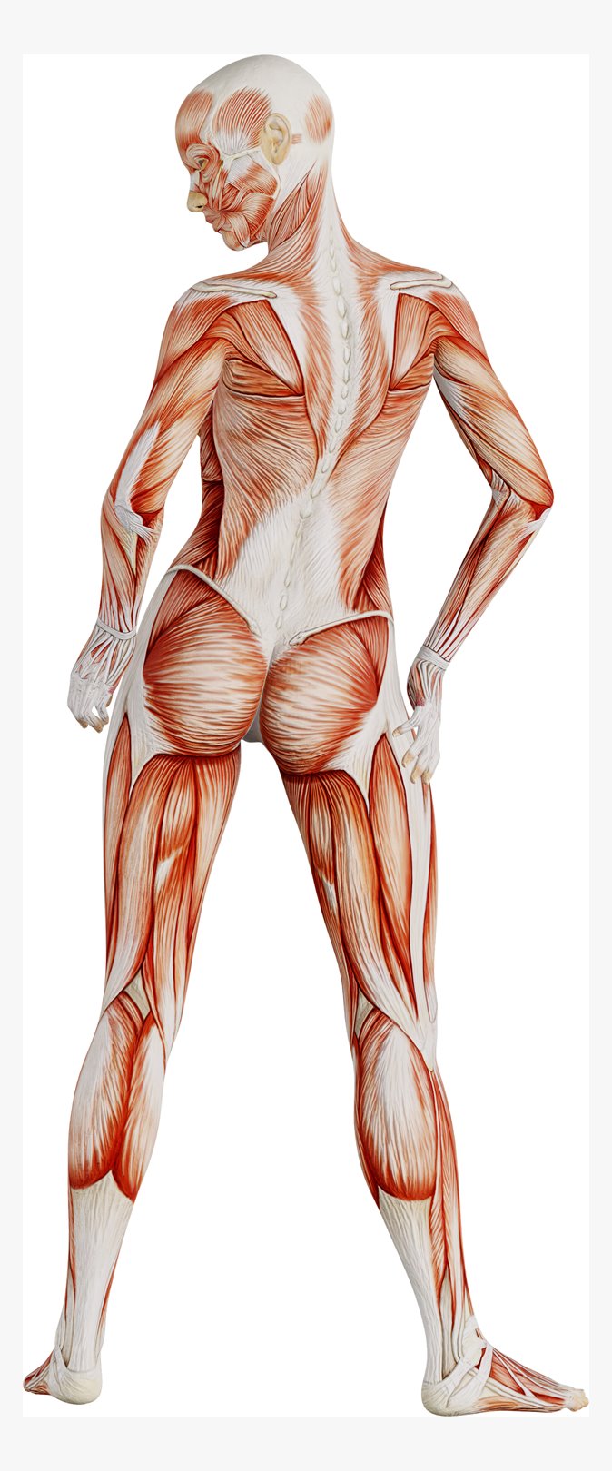
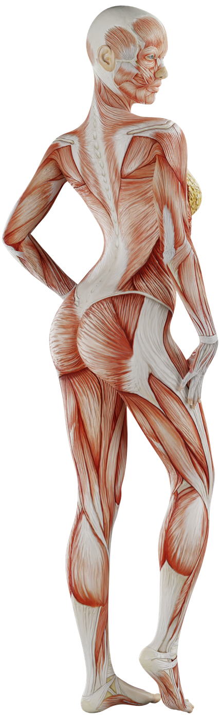
The very figure from the interactive: full height, seen from behind. Trapezius, deltoids, latissimus dorsi, erector spinae, glutes, hamstrings, calves — the whole superficial layer. A click on an area opens the card for that muscle.
Trapezius (m. trapezius)
Deltoid (m. deltoideus)
Latissimus dorsi (m. latissimus dorsi)
Erector spinae (m. erector spinae)
Thoracolumbar fascia (fascia thoracolumbalis)
Glutes (mm. glutei)
About pressure: “if you can breathe, it is fine”
Deep tissue massage is not “put up with it if it hurts”. The working level of discomfort is around 6-7 out of 10: clearly felt, definitely not pleasant, but you are breathing evenly, not holding your breath and not bracing. The moment you have to endure it, the body starts defending itself, the muscle gets stiffer — and the work loses its point. Say “less” at any moment; that does not interrupt the session, that is how you steer it.
A good complaint sounds like this: where, how long, what makes it worse, what makes it easier, what you did the day before. Four sentences, and half the search is already done.
Ale
Angles
The same body from six sides
A muscle is hard to understand from a single picture: in different positions the layers separate and you see what standing upright covers. These frames are used in the atlas and on the service pages — look at how differently one and the same back can read.
Muscles in focus
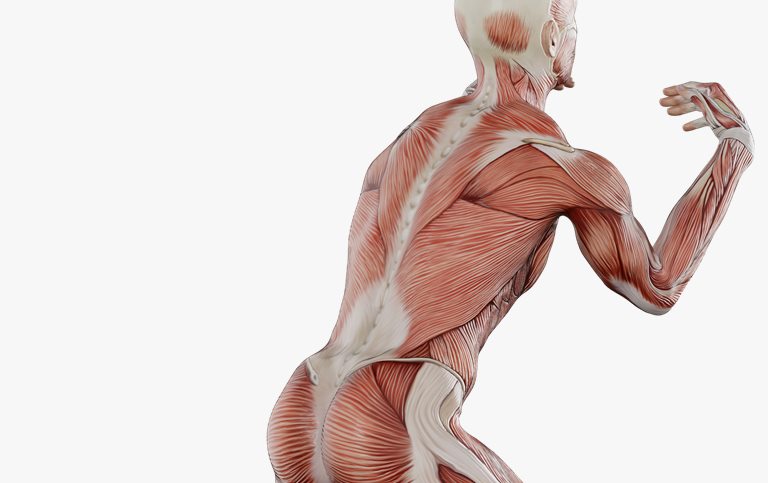
From behind, arms raised
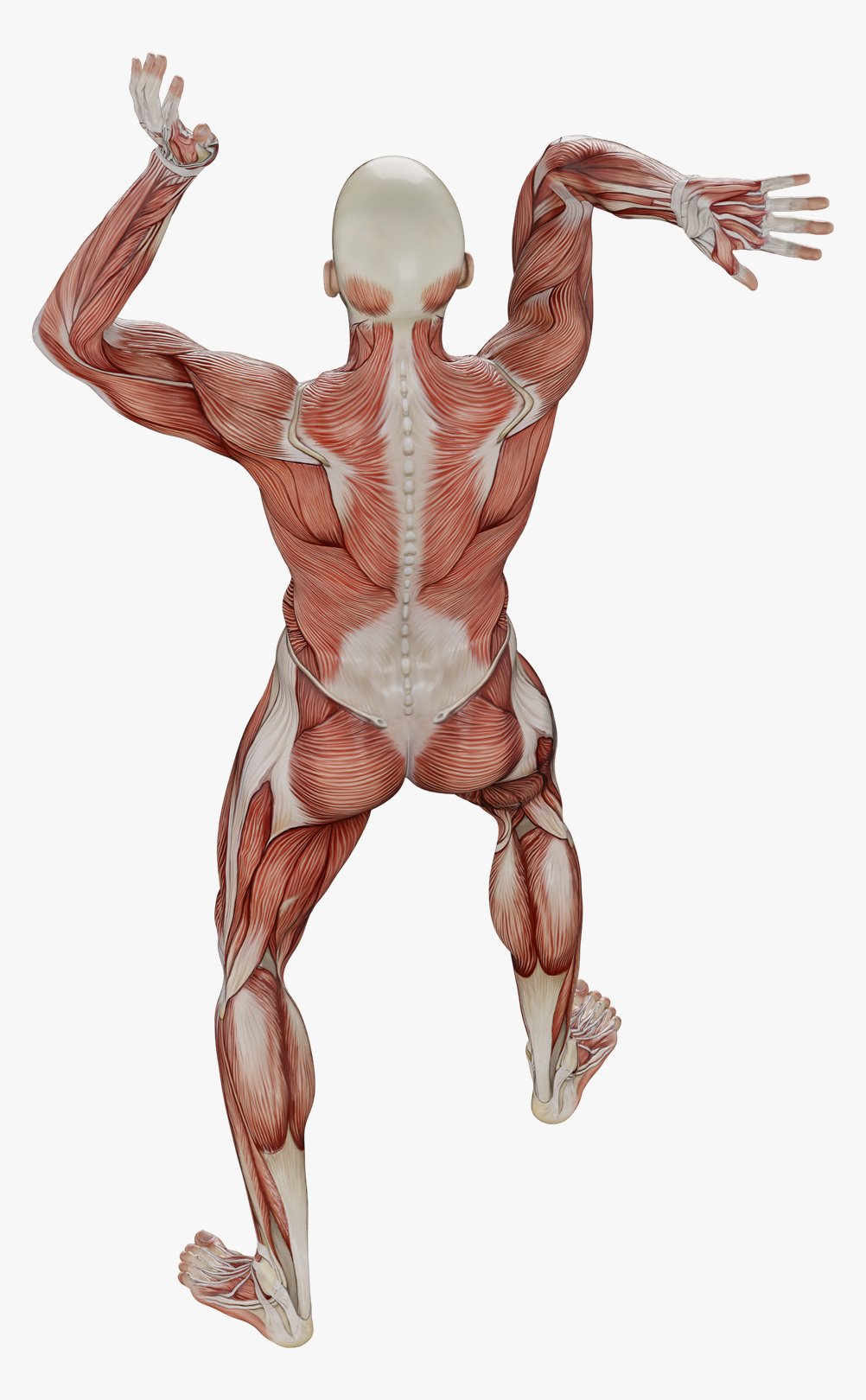
The base frame of the atlas. The trapezius in full — from the neck to mid-back, the deltoids, latissimus dorsi, erector spinae, thoracolumbar fascia, glutes, hamstrings and calves. Raised arms rotate the shoulder blade and open up what it covers in an ordinary standing position.
Trapezius (m. trapezius)
Deltoid (m. deltoideus)
Latissimus dorsi (m. latissimus dorsi)
Erector spinae (m. erector spinae)
Thoracolumbar fascia (fascia thoracolumbalis)
Glutes (mm. glutei)
Muscles in focus
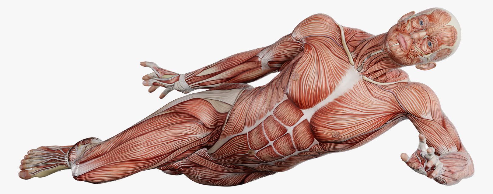
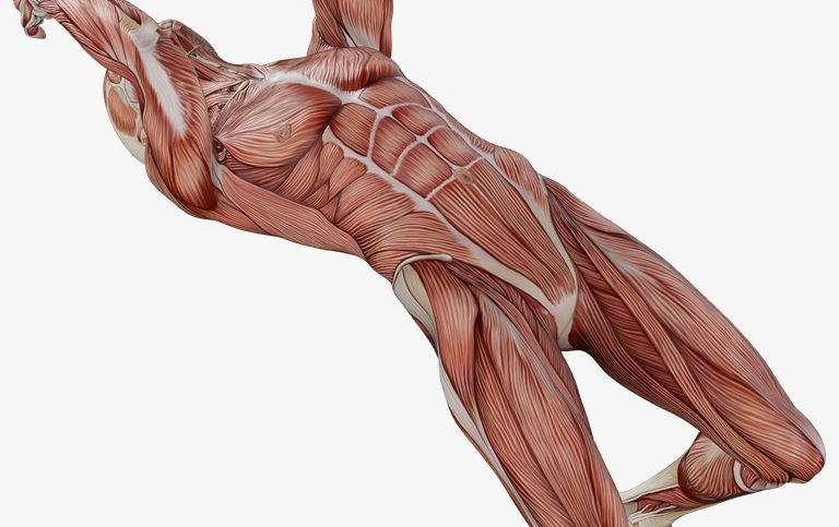
From the front, reclining
The front half, the one nobody thinks about when their back hurts: pectoralis major, rectus abdominis, the obliques, serratus anterior (m. serratus anterior), quadriceps, biceps brachii. Shoulders that have drifted forward are pulled from the front — while the ache turns up behind.
Pectoralis major (m. pectoralis major)
Rectus abdominis (m. rectus abdominis)
External oblique (m. obliquus externus abdominis)
Serratus anterior (m. serratus anterior)
Quadriceps (m. quadriceps femoris)
Biceps brachii (m. biceps brachii)
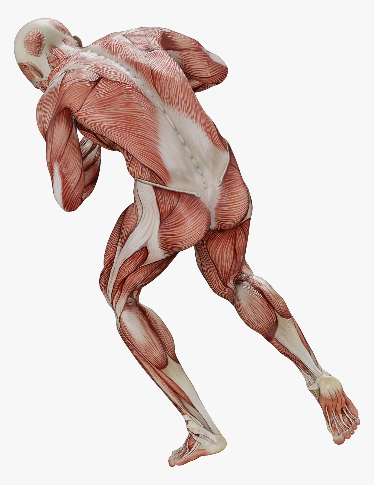
Muscles in focus
Forward bend
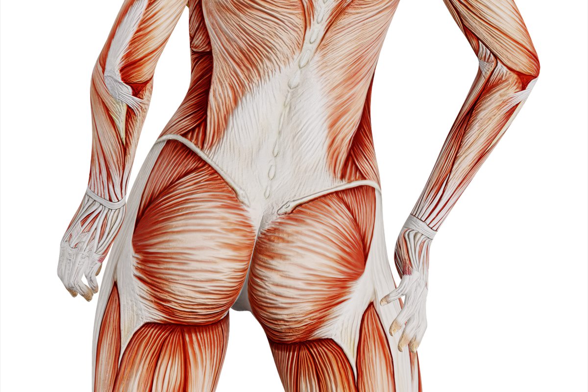
The thoracolumbar fascia up close — the dense diamond at the base of the back through which the lower back is tied to the lats and the glutes. In a bend it goes taut, and you can see why the lower back is almost never worked on its own, without the pelvis and the hamstrings.
Thoracolumbar fascia (fascia thoracolumbalis)
Latissimus dorsi (m. latissimus dorsi)
Glutes (mm. glutei)
Hamstrings (mm. ischiocrurales)
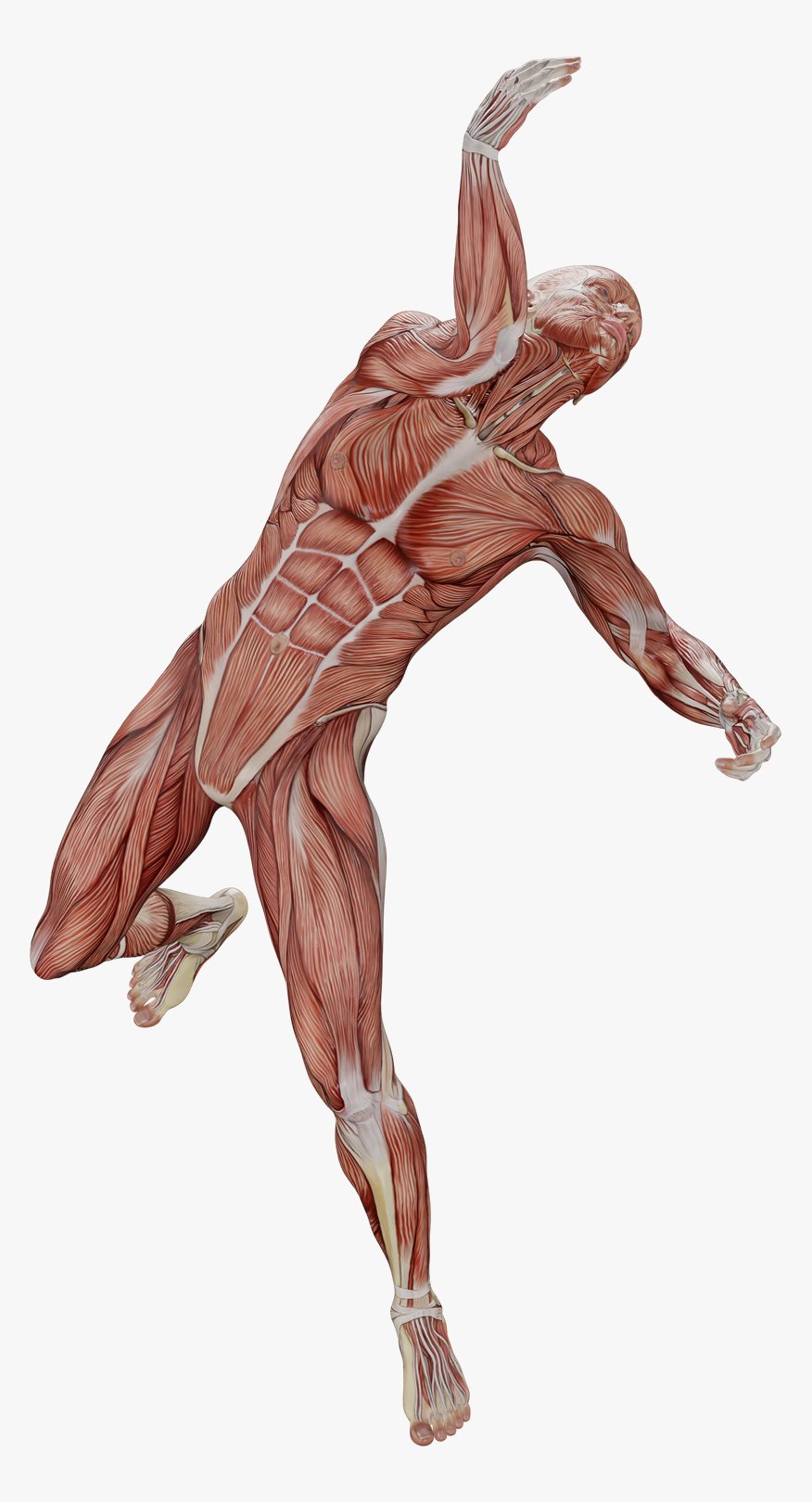
Muscles in focus
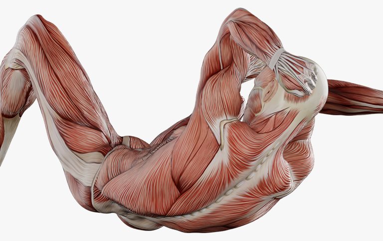
Torso rotation
External oblique, serratus anterior, rectus abdominis, quadriceps. Rotation is the one movement that most often “will not go” in people who sit at a desk, and it is tested before and after a session.
External oblique (m. obliquus externus abdominis)
Serratus anterior (m. serratus anterior)
Rectus abdominis (m. rectus abdominis)
Quadriceps (m. quadriceps femoris)
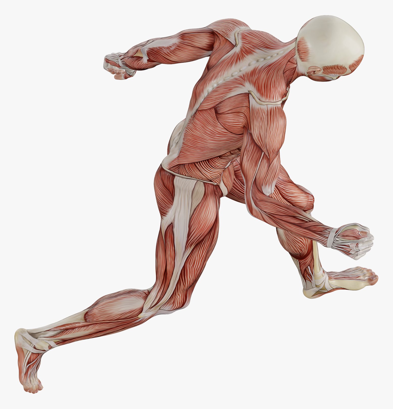
Muscles in focus
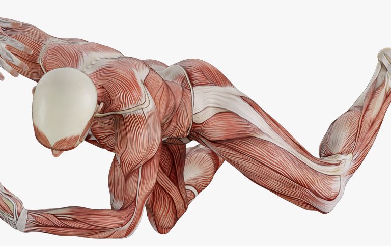
Running stride
Glutes, hamstrings, the calf and the Achilles tendon at the moment of load. Here you can clearly see the chain that a sports and recovery session takes apart: pelvis, thigh and shin work as a single line.
Glutes (mm. glutei)
Hamstrings (mm. ischiocrurales)
Gastrocnemius (m. gastrocnemius)
Achilles tendon (tendo calcaneus)
Muscles in focus
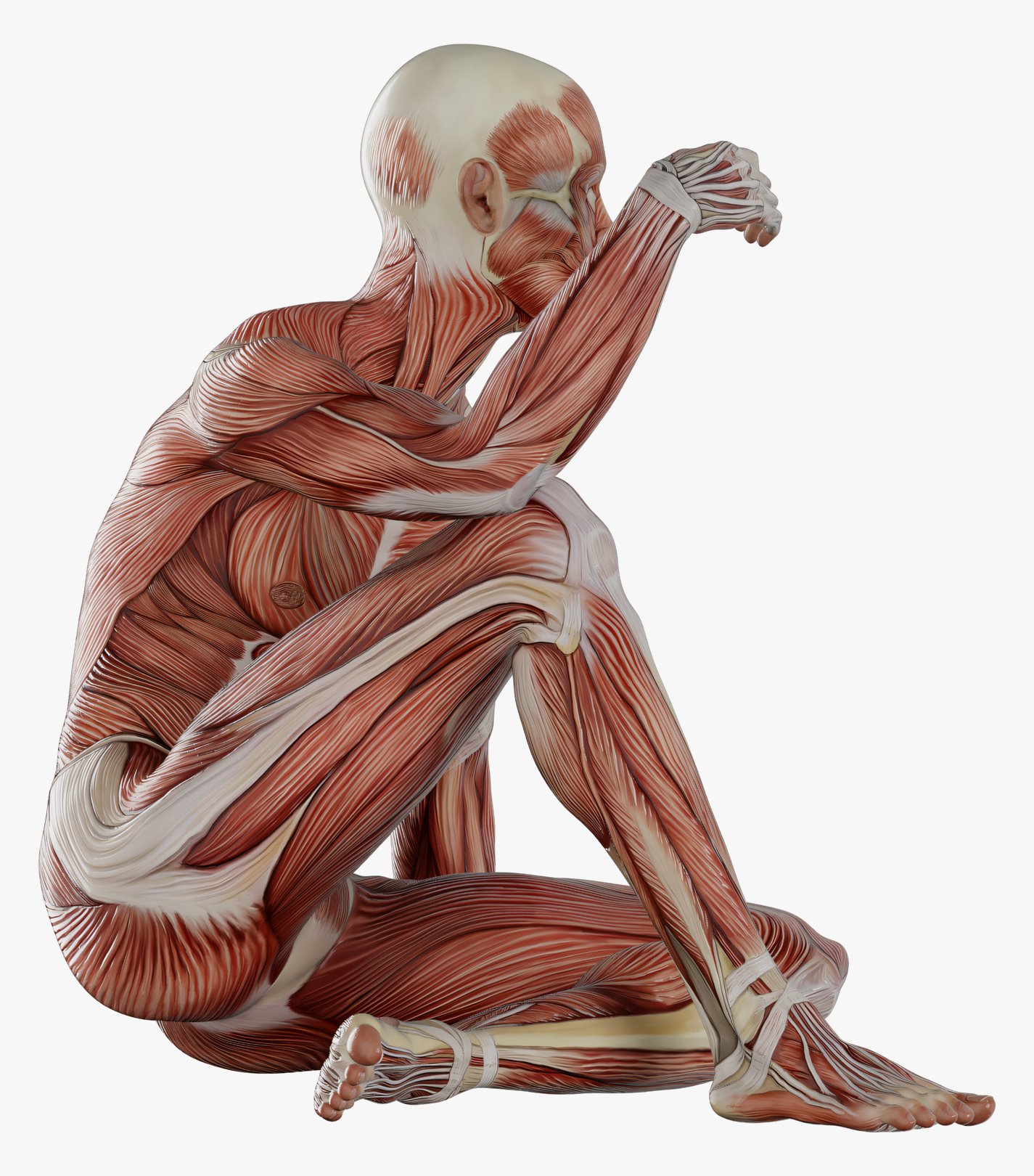
Seated, arms around the knees
Neck and upper trapezius, deltoid, erector spinae, glutes, calf. A position in which the superficial layer of the back is under tension — and under it hide the short suboccipital muscles, which you reach with your hands rather than with your eyes.
Upper trapezius (m. trapezius, pars descendens)
Deltoid (m. deltoideus)
Erector spinae (m. erector spinae)
Glutes (mm. glutei)
Gastrocnemius (m. gastrocnemius)
Referred pain
Where the point is — and where it refers
Muscle pain often shows up somewhere other than the overloaded muscle. Pick a point on the figure — the area it usually refers to lights up, and a description opens: which muscle it is, what sets it off and what helps.
Front
Back
1trigger point — tap it
main referral zone
spillover zone — not in everyone
Points
1Point · Back
Upper trapezius
m. trapezius, pars descendens
Where the point is
Middle of the shoulder ridge, between the neck and the shoulder.
Where it refers
Back and side of the neck, behind the ear, temple; sometimes the angle of the jaw.
What usually sets it off
Screen below eye level, phone held by the shoulder, bag on one side.
What helps
Ischaemic compression and a gentle side stretch; screen at eye level, a headset instead of the shoulder.
Zone shown: Front · Back
If the “migraine” came on suddenly and is the worst of your life, or comes with slurred speech, weakness or numbness on one side of the body, it is an emergency.
2Point · Back
Levator scapulae
m. levator scapulae
Where the point is
Above the upper corner of the shoulder blade, at the back of the base of the neck.
Where it refers
The angle between the neck and the shoulder; along the inner edge of the shoulder blade.
What usually sets it off
Head turned to a side monitor, a high pillow, a bag on the shoulder.
What helps
Compression at the upper corner of the shoulder blade, a “nose to armpit” stretch; the monitor straight ahead.
Zone shown: Back
A neck that will not turn after an injury, or with numbness in the arm, is a reason to see a doctor, not to book a massage.
3Point · Back
Suboccipital muscles
mm. suboccipitales
Where the point is
Under the edge of the skull at the back, deep, either side of the midline.
Where it refers
A band from the back of the head through the temple to the eye.
What usually sets it off
Chin poking towards the screen, glasses with the wrong focus zone, long reading lying down.
What helps
Gentle sustained compression under the skull, a “double chin” nod; screen and glasses set for the working distance.
Zone shown: Front · Back
A headache with fever and a stiff neck is an emergency.
4Point · Front
Sternocleidomastoid
m. sternocleidomastoideus
Where the point is
Front and side of the neck: the cord from the collarbone and breastbone up behind the ear.
Where it refers
Forehead, around the eye, the ear, the top of the head.
What usually sets it off
Head tilted back to a high screen, sleeping face down with the head turned, a lingering cough.
What helps
A pincer grip, with no pressure deep into the neck; a head position without a constant turn.
Zone shown: Front · Back
Dizziness, ringing in the ear and loss of balance are for a doctor to check first.
5Point · Front
Scalene muscles
mm. scaleni
Where the point is
Side of the neck, above the collarbone.
Where it refers
Chest, shoulder, outer side of the arm, forearm, thumb and index finger; the inner edge of the shoulder blade.
What usually sets it off
Upper-chest breathing, stress, coughing, carrying heavy loads, driving.
What helps
Careful finger work, belly breathing; less hanging on the steering wheel and carrying heavy loads in one hand.
Zone shown: Front · Back
Numbness and weakness in the hand, or numb fingers at night — see a neurologist.
6Point · Back
Infraspinatus
m. infraspinatus
Where the point is
On the shoulder blade, below its spine.
Where it refers
Deep into the front of the shoulder and down the front and outer side of the arm.
What usually sets it off
Using a mouse at arm’s length, tennis and swimming, sleeping on the side with the arm under the head.
What helps
Compression on the shoulder blade, not on the shoulder where it hurts; the mouse closer to the body.
Zone shown: Front
Night pain in the shoulder, weakness when raising the arm, or an injury — see a doctor first.
7Point · Back
Quadratus lumborum
m. quadratus lumborum
Where the point is
Side of the lower back, between the lowest rib and the iliac crest.
Where it refers
Sacroiliac joint, buttock, greater trochanter — the outer side of the hip.
What usually sets it off
Bending with a twist, standing on one leg for long, a wallet in the back pocket, legs of different length.
What helps
Work lying on the side, a side stretch; lifting from the floor without twisting.
Zone shown: Back
Low back pain with fever, night pain or bladder problems — see a doctor.
8Point · Back
Gluteus medius
m. gluteus medius
Where the point is
Below the iliac crest, in the upper outer part of the buttock.
Where it refers
The lower back along the iliac crest, the sacrum.
What usually sets it off
Sleeping on the side, running on cambered ground, standing “on one hip” for long.
What helps
Compression below the iliac crest, strengthening the hip abductors.
Zone shown: Back
Pain that runs below the knee together with numbness is not this route.
9Point · Back
Gluteus minimus
m. gluteus minimus
Where the point is
Deep under the gluteus medius, in the outer part of the buttock.
Where it refers
Outer and back of the thigh and lower leg, sometimes to the ankle — “like sciatica”.
What usually sets it off
Long sitting, sleeping on that side, running on cambered ground.
What helps
Slow deep compression through the gluteus medius; getting up every 40–50 minutes.
Zone shown: Front · Back
Numbness, a weak foot, pain in a narrow strip, worse with coughing — signs of a nerve root, see a neurologist.
10Point · Back
Soleus
m. soleus
Where the point is
Lower part of the back of the leg, under the gastrocnemius.
Where it refers
Back and underside of the heel, less often the sacral area.
What usually sets it off
New shoes, hard surfaces, a sharp jump in running volume.
What helps
Work on the calf with the knee bent, stretching with the knee bent; build volume gradually.
Zone shown: Back
A swollen calf that is red, warm and firm may be a thrombosis: massage is contraindicated, see a doctor at once.
The map is a working model, not a diagnosis. Numbness, weakness, shooting pain — see a doctor, not a massage table.
Patterns after Travell & Simons, “Myofascial Pain and Dysfunction: The Trigger Point Manual”. Each zone is drawn on one side of the body for clarity, although it can occur on both; in a given person the zone may be smaller or shifted. A diagram, not a measurement.
Problem areas
The four bundles people come in with most often
Muscles rarely get tired one at a time — they work in bundles. So a session takes apart not a “point” but a bundle: the area, its neighbours and whatever is pulling on it from the other side. Here are the four most common ones.
The “eight hours at a laptop” bundle. People complain about the neck, but the neck is rarely the short part: on top the upper trapezius is overloaded from holding the shoulder blade for hours, and from the front pectoralis minor pulls — you cannot see it on a superficial render, it lies under pectoralis major. The work always goes on both sides: the back half alone buys you a day or two.
The “seated lower back” bundle. The ache is usually to one side and a little above the pelvis — where the quadratus lumborum (QL) sits: it runs deeper and is not visible in this frame, and it gets confused with the erector spinae constantly, although they are different muscles doing different jobs. Almost always the bundle also includes the glutes and hamstrings, which are clearly visible: a pelvis that does not extend hands the movement over to the lower back.
The “it pulls from the buttock downwards” bundle. Under gluteus maximus lies a deep layer that includes the piriformis — you cannot see it on a render of the superficial layer, but it is often the one that turns out to be tender on palpation. An important boundary: if numbness or tingling runs down the leg, or the foot is getting weaker, the session is cancelled and you go to a doctor.
The bundle people skip. When the back aches behind, it is often being pulled from the front: the chest and the front of the thigh are shortened from sitting. Along the outside of the thigh — it barely makes it into this frame — runs the iliotibial band (ITB): a fascial strap, not a muscle, and you cannot “stretch” it with massage. The work goes into what weaves into it: the tensor fasciae latae (TFL) and vastus lateralis.
Short answers to the things people most often write after their first look at this page.
No. The atlas is a convenience, not an entrance exam. Four facts are enough: where you feel it, how long it has been going on, what makes it worse and what makes it easier. Everything else is my job: to find it, check it by hand and confirm it against your own sensations. You will pick up the muscle names along the way if you want to, simply because I say out loud what I am doing and why.
No, and that matters. A matching description is a good hypothesis and an excellent starting point for a conversation, but not a conclusion about the cause. Very different things produce similar sensations, and some of them have nothing to do with massage at all. I do not make diagnoses and do not prescribe treatment — I work with muscles and the way they move. Send me your version and we will test it during the session rather than take it on faith.
Because of referred pain: a dense spot in a muscle can produce a sensation away from itself, sometimes ten centimetres away. The second mechanism is compensation: the area that aches is often simply doing the work of a neighbour that is not moving. The classic case is a lower back taking over the movement of a pelvis that will not extend. So the hands almost always go wider than the pain does, and I explain why they are going exactly there.
Within reason — yes, and I usually show a couple of things myself at the end of a session. The rules are simple: soft tissue, not bones and not joints; never along the spine; not on the abdomen and not on the front of the neck; a minute and a half to two minutes per area, not twenty; breathe evenly. If things got worse after working on yourself, or the pain holds for more than a day, stop and write to me. And a roller is no substitute for a doctor when the symptom is new and unclear.
When there is numbness, tingling, weakness in an arm or a leg, loss of sensation, a shooting pain after which the limb will not obey; with an injury, swelling, fever, signs of inflammation; with pain that wakes you at night and does not depend on position; with unexplained weight loss. Separately: thrombosis and thrombophlebitis, cancer (only in agreement with your treating doctor), anticoagulant therapy, uncontrolled hypertension, pregnancy — these need a specialist in that field. The full list is in the contraindications section. I will say so plainly if the case is not mine.
A relaxed standing view from behind: the neutral reference against which the tilt of the shoulders and pelvis is compared before a first session. Trapezius (m. trapezius) · Latissimus dorsi (m. latissimus dorsi) · Erector spinae (m. erector spinae) · Glutes (mm. glutei)
Found your area? Come in with it
Bring one sentence with you: where it pulls, how long it has done so, what makes it worse. I will check it by hand, show you the difference in movement before and after, and tell you honestly if this is not for me. Sessions at the studio at Balmes 200, plus visits to homes, hotels and offices — Area Metropolitana de Barcelona.